By Nauman F. Manzoor, M.D. * and Viral Tejani, Au.D., Ph.D. *
Department of Otolaryngology-Head and Neck Surgery, University Hospitals / Case Western Reserve University School of Medicine
*Co-authors with equal contribution to the write-up
Patient background
An 88 year old female was referred for bilateral progressive sensorineural hearing loss and remote history of left- sided Meniere’s Disease. She no longer had any episodic vertigo but did endorse mild imbalance. She was using bilateral traditional amplification but over the last year had noticed decline in speech understanding. Based on comprehensive audiometric testing, she was a cochlear implant candidate in both ears. Vestibular testing showed left-sided weakness and hence this ear was selected for cochlear implantation. Pre-operative computed tomography (CT) imaging was normal with no cochlear or labyrinthine abnormality (images below). Normal mastoid anatomy and facial nerve course was appreciated. Prior magnetic resonance imaging (MRI) scan was also negative for any retrocochlear lesion.


Device selection / Electrophysiology background
In all standard cochlear implant cases, we use CI632 Slim Modiolar Electrode (SME) for modiolar proximity and good cochlear coverage. While clinical studies indicate a low tip fold-over rate1-5 with the SME, our institution still routinely confirm device placement via electrophysiologic measures and an intraoperative x-ray using a modified stenver view.
While effective, these imaging measures add to the surgical costs, expose patients to radiation (albeit a small amount), and add to the surgery time3. Thus, electrophysiological measures of TransImpedance Matrix (TIM) have been previously proposed and has shown promise in identifying tip fold-over5-6. Essentially, the TransImpedance Matrix show how voltage is distributed along the electrode array when the cochlear implant is stimulated.
The TransImpedance Matrix tool is available in Custom Sound® and has also been incorporated into the recently created Nucleus® SmartNav platform. The SmartNav platform is an iPad based platform that wirelessly interfaces with the internal CI; it monitors insertion speed and conducts a “Placement Check” as well as impedance and AutoNRT recordings once the CI is inserted. The placement check is a TransImpedance Matrix recording.
Surgical course
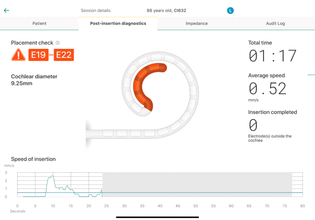
A standard mastoidectomy was performed and a facial recess approach was used. Round window anatomy was normal, and an extended round window approach was utilized. The electrode array was deployed in a standard manner keeping a co-planar trajectory to the basal turn. After placement, the SmartNav System was used to conduct a Placement Check. As shown by the screenshot below, the placement check indicated a tip fold-over at electrodes 19-22.

To verify the Placement Check results and obtain more detailed measure, a TransImpedance Matrix recording using Custom Sound was performed.* These results are plotted in the below figure. The left axis shows the recording electrode, while the bottom axis shows the stimulating electrode. In a normal TransImpedance Matrix plot, a dark black / red diagonal line from the lower left hand corner to the upper right hand corner is expected, which shows normal voltage distribution. However, the below figure indicates abnormal voltage spread at the apical electrodes starting around E15-22 (see lower left hand corner plot). For example, when looking at stimulating electrode 22, one can see two peaks (as signified by the red points at recording electrode 22 and recording electrode 15/16). This signifies that electrodes 15 and 22 are very close to one another due to the high voltage recordings.
*TIM is only available in Custom Sound® EP and requires a regional key and access code.

This was confirmed via an intraoperative X-ray. It does appear that there is fold-over starting and that electrode 22 and electrodes 15/16 are very close to one another.

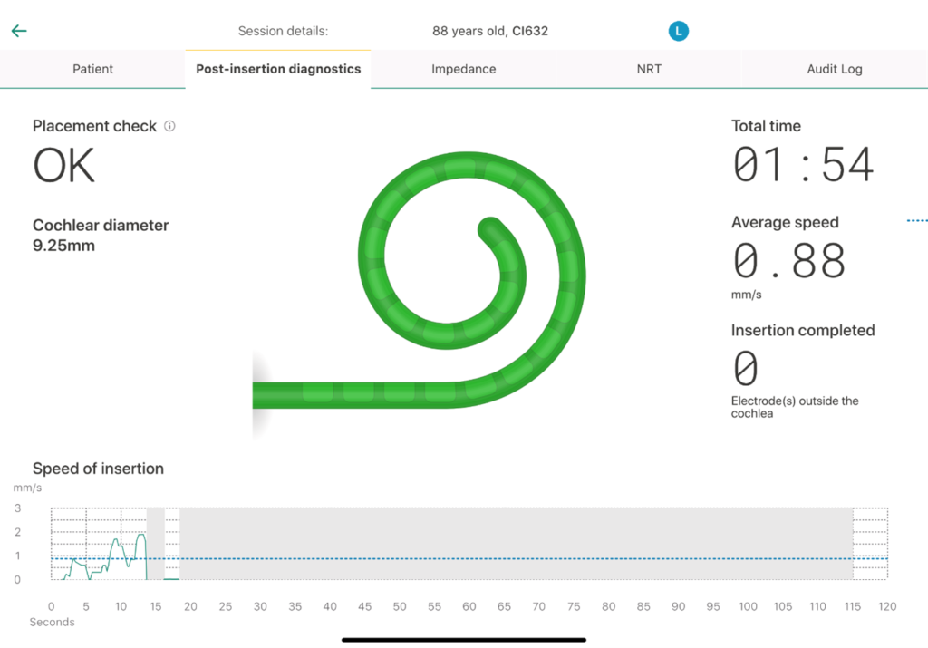
The electrode was removed, reloaded into the sheath, and the array was reinserted without incident. SmartNav Placement Check was normal.

The TransImpedance Matrix recording was also normal (see below figure). As previously stated, in a normal TransImpedance Matrix plot, we expect a dark black / red diagonal line from the lower left hand corner to the upper right hand corner.

Intraoperative X-ray was also normal.

Conclusions
This case study provides proof-of-concept regarding the utility of TransImpedance Matrix recordings (as embedded in SmartNav or as separately available in Custom Sound) in evaluating cochlear implant placement. Given the low tip-foldover rate reported across the literature for the SME array1-5, large scale studies are needed to generate a sufficient patient sample size to verify the sensitivity and specificity of TransImpedance Matrix recordings in identifying tip-foldovers and compare these metrics to the sensitivity of intraoperative imaging.
Get an inside look on the SmartNav System here!
About the authors:

Dr. Manzoor is a board-certified otolaryngologist and head & neck surgeon with fellowship training in otology-neurotology and lateral skull base surgery. He has expertise in the diagnosis and treatment of a wide range of otologic, neurotologic and skull base conditions. He completed his otolaryngology training at University Hospitals Cleveland Medical Center and completed his fellowship training at Vanderbilt University Medical Center. He is particularly passioned about hearing rehabilitation and strives to deliver cutting edge treatment options for patients with hearing disorders. He is also an avid researcher with 60 peer reviewed publications related to otologic-neurotologic disorders and outcomes.

Viral Tejani, AuD, PhD, is a Cochlear Implant Audiologist and Assistant Professor of Otolaryngology. He sees cochlear implant patients in the clinic and is also involved with both NIH-funded and industry-funded research. His particular expertise/passions are in cochlear implant psychophysics, electrophysiology, electric-acoustic stimulation, as well as improving outcomes of cochlear implantation. He completed his AuD at University of Maryland and his PhD at University of Iowa, where he remained as an Otolaryngology faculty member until relocating to Cleveland.
References
- Aschendorff, A., Briggs, R., Brademann, G., Helbig, S., Hornung, J., Lenarz, T., Marx, M., Ramos, A., Stöver, T., Escudé, B., & James, C. J. (2017). Clinical investigation of the Nucleus Slim Modiolar Electrode. Audiology & neuro-otology, 22(3), 169–179. https://doi.org/10.1159/000480345
- Friedmann, D. R., Kamen, E., Choudhury, B., & Roland, J. T., Jr (2019). Surgical Experience and Early Outcomes With a Slim Perimodiolar Electrode. Otology & neurotology : official publication of the American Otological Society, American Neurotology Society [and] European Academy of Otology and Neurotology, 40(3), e304–e310. https://doi.org/10.1097/MAO.0000000000002129
- Klabbers, T. M., Huinck, W. J., Heutink, F., Verbist, B. M., & Mylanus, E. (2021). Transimpedance Matrix (TIM) Measurement for the Detection of Intraoperative Electrode Tip Foldover Using the Slim Modiolar Electrode: A Proof of Concept Study. Otology & neurotology : official publication of the American Otological Society, American Neurotology Society [and] European Academy of Otology and Neurotology, 42(2), e124–e129. https://doi.org/10.1097/MAO.0000000000002875
- Shaul, C., Weder, S., Tari, S., Gerard, J. M., O’Leary, S. J., & Briggs, R. J. (2020). Slim, Modiolar Cochlear Implant Electrode: Melbourne Experience and Comparison With the Contour Perimodiolar Electrode. Otology & neurotology : official publication of the American Otological Society, American Neurotology Society [and] European Academy of Otology and Neurotology, 41(5), 639–643. https://doi.org/10.1097/MAO.0000000000002617
- Shew, M. A., Walia, A., Durakovic, N., Valenzuela, C., Wick, C. C., McJunkin, J. L., Buchman, C. A., & Herzog, J. A. (2021). Long-term Hearing Preservation and Speech Perception Performance Outcomes With the Slim Modiolar Electrode. Otology & neurotology : official publication of the American Otological Society, American Neurotology Society [and] European Academy of Otology and Neurotology, 42(10), e1486–e1493. https://doi.org/10.1097/MAO.0000000000003342
- Zuniga, M. G., Rivas, A., Hedley-Williams, A., Gifford, R. H., Dwyer, R., Dawant, B. M., Sunderhaus, L. W., Hovis, K. L., Wanna, G. B., Noble, J. H., & Labadie, R. F. (2017). Tip Fold-over in Cochlear Implantation: Case Series. Otology & neurotology : official publication of the American Otological Society, American Neurotology Society [and] European Academy of Otology and Neurotology, 38(2), 199–206. https://doi.org/10.1097/MAO.0000000000001283
SmartNav offers a variety of measurements. These insights are one source of information and do not guarantee clinical outcomes.
Apple, the Apple logo, Apple Watch, FaceTime, Made for iPad logo, Made for iPhone logo, Made for iPod logo, iPhone, iPad Pro, iPad Air, iPad mini, iPad and iPod touch are trademarks of Apple Inc., registered in the U.S. and other countries. App Store is a service mark of Apple Inc., registered in the U.S. and other countries.


